What Causes Keratoconus?
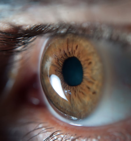
While some experts think patients are born with a certain predilection to keratoconus (and we actually have a genetic test that we offer at our practice to see if you have any of the keratoconus genetic factors), most agree that the only significant risk factor is rubbing of the eyes. Multiple episodes of eye rubbing exerted onto the cornea, can cause corneal deformation and thinning of the cornea, which then can lead to keratoconus. If you only take one thing away from this information—please avoid rubbing your eyes!
1 Belin MW, Lim L, Rajpal RK, Hafezi F, Gomes JAP, Cochener B. Corneal Cross-Linking: Current USA Status: Report From the Cornea Society. Cornea. 2018 Oct;37(10):1218-1225. doi: 10.1097/ICO.0000000000001707. Erratum in: Cornea. 2019 Oct;38(10):e49. PMID: 30067537.
2 Chatzis, Nico, and Farhad Hafezi. “Progression of Keratoconus and Efficacy of Corneal Collagen Cross-Linking in Children and Adolescents.” Journal of Refractive Surgery, vol. 28, no. 11, 11 Oct. 2012, pp. 753–758., https://doi.org/10.3928/1081597x-20121011-01.
3 Kreps EO, Claerhout I, Koppen C. Diagnostic patterns in keratoconus. Cont Lens Anterior Eye. 2021 Jun;44(3):101333. doi: 10.1016/j.clae.2020.05.002. Epub 2020 May 21. PMID: 32448765.
4 Kalra, Nidhi, et al. “Posterior Chamber Phakic Intraocular Lens Implantation for Refractive Correction in Corneal Ectatic Disorders: A Review.” Journal of Refractive Surgery, vol. 37, no. 5, 1 May 2021, pp. 351–359. https://doi.org/10.3928/1081597x-20210115-03.
5 Ramin S, Sangin Abadi A, Doroodgar Fet al.. Comparison of visual, refractive and aberration measurements of INTACS versus Toric ICL lens implantation; a four-year follow-up. Med Hypothesis Discov Innov Ophthalmol. 2018; 7(1):32–39.29644243.
6 Nattis, Alanna S. DO, FAAO; Rosenberg, Eric D. DO, MSE; Donnenfeld, Eric D. MD One-year visual and astigmatic outcomes of keratoconus patients following sequential crosslinking and topography-guided surface ablation: the TOPOLINK study, Journal of Cataract and Refractive Surgery: April 2020 – Volume 46 – Issue 4 – p 507-516. doi: 10.1097/j.jcrs.0000000000000110
7 Sakla, Hani F., et al. “Visual and Refractive Outcomes of Toric Implantable Collamer Lens Implantation in Stable Keratoconus after Combined Topography-Guided PRK and CXL.” Journal of Refractive Surgery, vol. 37, no. 12, 1 Dec. 2021, pp. 824–829., https://doi.org/10.3928/1081597x-20210920-02.


